Research Associate, Dalio Institute of Cardiovascular Imagining, Department of Radiology at Weill Cornell Medicine
New York-Presbyterian
Overview
At the Dalio Institute of Cardiovascular Imaging, Sun-Joo Jang and his team have undertaken the task of using a mix of augmented and virtual reality to aid in cardiology procedures.
In this presentation, Jang describes the process that he and his team used to build a deep learning algorithm to create accurate and real-time three-dimensional models for use during operations.
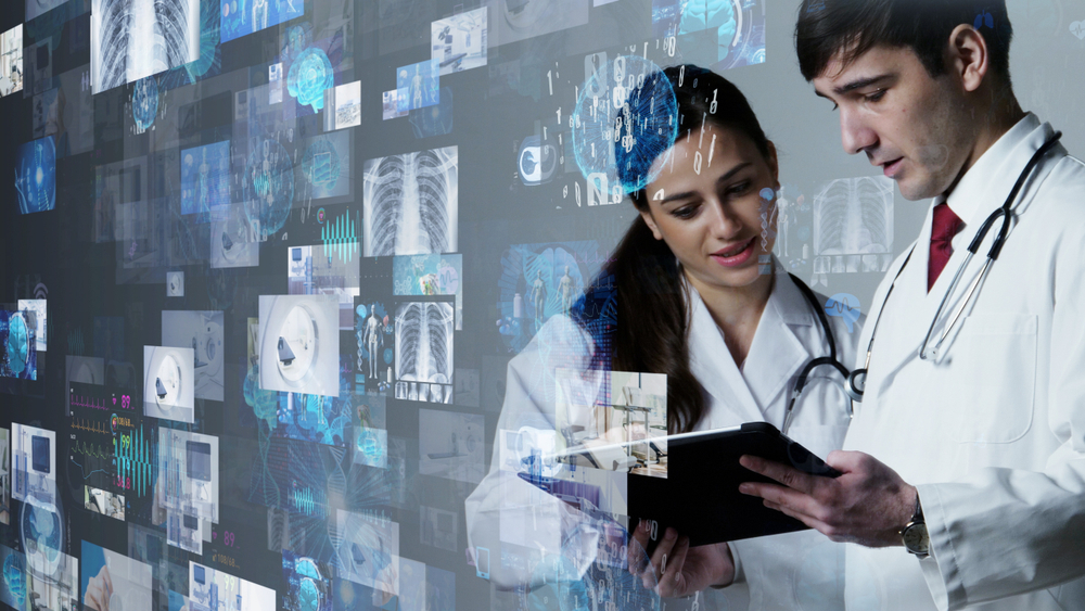
Technological advances continue to aid the medical community in their capacity to treat an increasing array of afflictions. Now, medical imaging has begun to take the next revolutionary step with the help of algorithms that allow medical professionals the ability to virtually step inside procedures being performed.
As Jang describes, virtual reality can be thought of as a reality in which you need to view through some sort of optics (usually glasses) while augmented reality is able to create a digital display that is separate from the real physical environment. At Dalio Institute of Cardiovascular Imaging, Jang and team are using a combination of these two to create a mixed reality where generated holograms can be interacted with in a three-dimensional virtual environment.
The benefits to using this combination of technology and machine learning can lend to substantial improvements in the ability for cardiologists to perform procedures effectively and efficiently. Often times when procedures are difficult or have a risk of serious complications additional preparation and staff are needed during the procedure. Being able to create these three-dimensional models to study prior to the procedure and then use them in real time during the procedure can greatly increase the success of procedures while at the same time decreasing the supporting cast cardiologists require.
“Education wise, understanding the anatomy has higher potential and mixed reality will play a much better role teaching future generations.”
Additionally, this mixed reality imaging is an excellent tool for teaching. Where more experienced cardiologists can tell where they are exactly positioned, less experienced or new cardiologist may have a difficult time translating the traditional two-dimensional CAT scans and X-rays into the real three-dimensional world. The mixed reality imaging aids in bridging this gap between the two-dimensional and three-dimensional worlds that often separates text book learning and real-world application.
Deep learning algorithms are similar to neural networks in the way that they are structured where data gets passed back and forth within an algorithm that attempts to optimize results. Where deep learning differs from neural networks is the amount of structure that is provided to the data set fed in to the modeling process and the expectations of the results that the model is told to look for. The data sets for deep learning models are unstructured and the targeted outputs are not defined. In this manner, the algorithm is allowed to come up with trends and expected outcomes free of human bias or persuasion.
Like neural networks, the data still needs to be trained, labelled, and validated. The challenge that Jang and team faced was being able to find consistent points of reference within the data set for which to build reliable models off of. First, external markers were placed along the spine to help define spaces. Next, X-ray images from many different angles were then used to determine proper marker locations and aid in filtering out shadows and other extraneous aspects in images.
Using this method an algorithm and model was created with over 99% accuracy that could place a catheter within 1mm of true position in three-dimensional space. As hearts are all shaped differently a flexible algorithm is required in order for catheters to be accurately guided into different spaces of the heart.
Sun-Joo Jang and his team now have the ability to bring a three-dimensional model of cardiovascular procedures in to any environment to the benefit of students or working professionals. For the purposes of education or being able to expertly navigate the intricacies of procedures, these models can be shared and utilized with precision and ease. Putting effort into the development and preparations of these models continue to advance the abilities that these medical professionals are able to provide and enhance the knowledge-base of all parties involved.